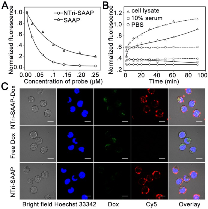
Figure 4.
Characterization of drug loading and release. (A) Drug loading capacity comparison of NTri-SAAP with SAAP based on fluorescence quenching of Dox. (B) Time-lapse fluorescence monitoring of Dox diffused from NTri-SAAP (solid line) or SAAP (dotted line) at 37 °C in cell lysate, 10% fetal bovine serum and PBS. (C) Confocal microscopy images of CEM cells after incubation with NTri-SAAP-Dox, free Dox, and pure NTri-SAAP (scale bar: 10 μm).

