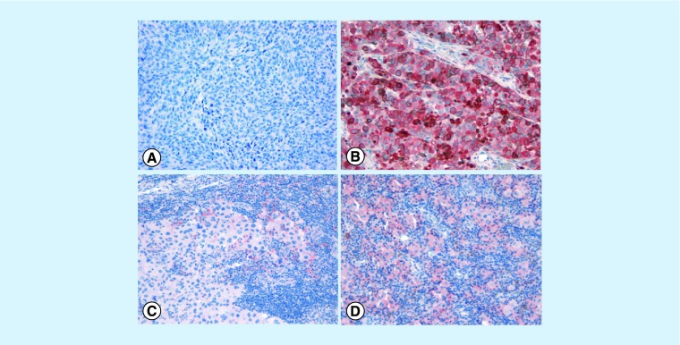
Figure 2. . PD-L1 status and immune cell infiltrates in the context of the melanoma microenvironment.
(A) PD-L1- melanoma in absence of immune cells; (B) strong and diffuse PD-L1 immunohistochemical staining in melanoma cells in absence of immune cells; (C) PD-L1 is expressed only focally in immune cells at the periphery of tumor aggregates but the tumor is PD-L1 negative; (D) PD-L1+ melanoma cells associated with prominent immune-cell infiltrate (original magnification ×20). The exact proportion of human melanomas that can be classified into these four categories is still an open issue although rough estimates suggest the following: PD-L1+/TILs+ 35%; PD-L1-/TILs- 40%; PD-L1+/TILs- 5% and PD-L1-/TILs+ 20% [18].

