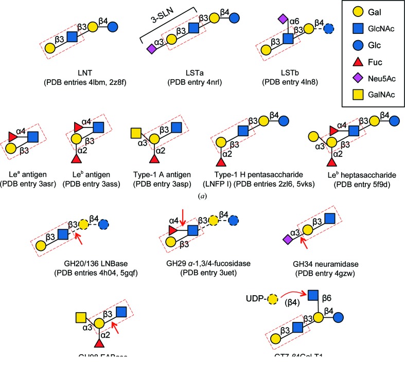
Figure 1.
Schematic representation of β-LNB-containing glycan structures in the PDB. (a) Glycan structures found in sugar-binding proteins and lectins. (b) Glycans observed in enzyme structures. The cleavage sites of the glyoside hydrolases and the transferase reaction site of the gycosyltransferase are indicated by red arrows. Disordered (and hence unmodelled) sugar moieties in sugar-binding protein structures, and substrate moieties additional to the crystallographic ligand of the enzymes, are shown with black dashed lines. The LNB unit is boxed with a red dashed line. For α-LNB-containing glycan structures, α-LNB disaccharide, LSTa and Lewis antigens were observed in a lectin from A. bisporus, in haemagglutinin from influenza virus and in VP1 (P domain) in norovirus, respectively.

