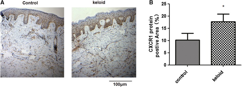
Fig. 3.
Measurement of CXCR1 protein expression in keloid tissues. Four pairs of tissue samples from keloid and surrounding regions were used to detect the protein levels of CXCR1 by immunohistochemistry. a Typical immunohistochemical images are shown. b Pooled data from a. The positive signaling with CXCR1 antibody staining was significantly stronger in keloid tissues than in the surrounding controls. The images shown are representative of four independent experiments. *P < 0.05

