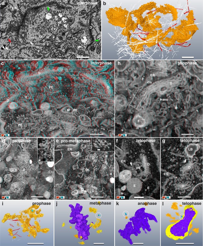
Fig. 6.
Golgi disintegration and reassembling. a SEM micrograph of an interphase Golgi apparatus. Single dictyosomes (D) are interconnected by shared single cisternae (green arrows). Abundant Golgi vesicles (V), primary endosomes (PE) and mitochondria (M) are located in between. MT (white arrows) and actin filaments (red arrow) interlace the Golgi. Scale bar 1 µm. a’ 3D-reconstruction of the interphase Golgi apparatus (of a) illustrates its 3D architecture. White lines representing a fraction of MTs, which interlace the Golgi-network. Actin fibers (red), circumjacent the cup-shaped Golgi. See Movie S3. Scale bar 1 µm. c Anaglyph image of the Golgi-network in interphase, revealing its three-dimensional architecture by interconnected dictyosomes (D). Several endosomes (E), vesicles (V) and mitochondria (M) are present in between. Scale bar 1 µm. d Golgi in early prophase, disintegrating synchronous into stacks and clouds of vesicles and vesicular–tubular clusters. High-resolution reveals that ERGICs (inset) represent the major fraction of the cloud, rather than single vesicles. Scale bar 1 µm; inset 100 nm. e Disintegrating Golgi in pro-metaphase: only rudimental dictyosomes (D), formed of vesicular–tubular clusters are present (inset). Scale bar 1 µm; inset 100 nm. f, g The Golgi reassembles in telophase. Single, separated dictyosomes form stacks of few cisternae. Typically, they are in direct contact to ER, primary endosome (PE) (f) and lipid bodies (g). Both, endosomes and lipid bodies have at least one or several connections to the ER. Scale bar 500 nm. h Anaglyph image of a dictyosome formed in telophase. Single dictyosomes (D) with characteristic cis- and trans-site are in contact to ER, lipid bodies and endosomes. ERGICs (framed area) are typically observed at the cis-site. Scale bar 1 µm. i–l Representative 3D-reconstructions of the entire Golgi apparatus (orange), chromatin (purple) and centrosomes (blue, encircled) at different mitotic stages. Scale bars 5 µm. i With onset of prophase the Golgi disintegrates rapidly into single dictyosomes, which collapse synchronous into clouds of vesicles and vesicular-tubular clusters. j After Golgi disassembly, several small, rudimentary dictyosomes and/or vesicle clusters are present in metaphase. k Only few rudimentary dictyosomes and vesicle clusters are still present in anaphase. l In telophase, groups of typical dictyosomes, sometimes interconnected, are visible in close proximity to the centrosome (circle) and on the opposite side of the nucleus

