Figure 5.
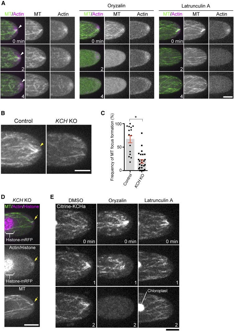
MT Focusing at the Apical Cell Tip Requires Actin Filaments and KCH.
(A) Formation of MT and actin foci (arrowheads) at the tip is mutually dependent. The tip of a caulonemal cell expressing GFP-tubulin (MT) and lifeact-mCherry (actin) was observed after oryzalin or latrunculin A treatment. Actin focal points were not maintained at the apical cell tip after oryzalin addition, whereas MT focal points were rarely observed after latrunculin A treatment.
(B) Persistent formation of the MT focus (arrowheads) at the apical cell tip is dependent on KCH.
(C) Frequency of MT focus formation. Images were acquired and analyzed every 3 s for 5 min. Bars and error bars represent the mean and se, respectively. Control, n = 15; KCH KO, n = 26. *P < 0.0001 (unpaired t test with equal sd, two-tailed). Observations were performed independently three times, and the data analyzed twice. The combined data are presented.
(D) Actin (lifeact-mCherry) and the transiently-formed MT focus (GFP-tubulin) in the absence of KCH (arrows).
(E) Actin-independent localization of Citrine-KCHa on MTs. Drugs were added at time 0, and images acquired at 0, 1, and 2 min are displayed. Note that signals derived from chloroplast autofluorescence are visible in some panels. Bars = 5 µm.

