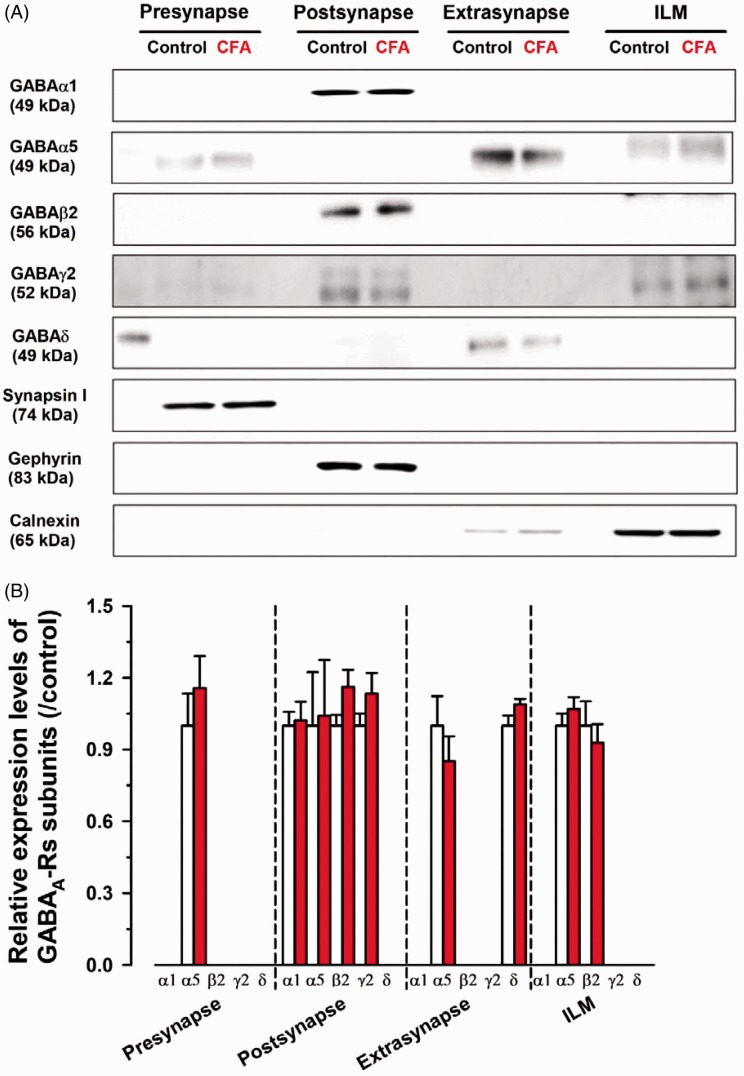
Figure 7.
The protein expression of GABAA receptor subunits was not altered in the ACC. (A) Western blotting analysis of GABAA receptor subunit expression. Synapsin I, gephyrin, and calnexin were used as presynaptic, postsynaptic (GABAergic), extrasynaptic, and ILM fraction markers, respectively. (B) Comparison of relative expression of GABAA receptor subunits between control and CFA in all fractions. In the presynaptic fraction of CFA group, the relative value of α5 was 1.15 ± 0.13 of control group. In the postsynaptic fraction of CFA group, the relative values of α1, α5, β2, and γ2 were 1.02 ± 0.08, 1.04 ± 0.23, 1.16 ± 0.07, and 1.13 ± 0.09 of control, respectively. In the extrasynaptic fraction of CFA group, the relative values of α5 and δ were 0.85 ± 0.10 and 1.10 ± 0.02 of control group. In ILM of CFA group, the relative values of α5 and β2 were 1.07 ± 0.05 and 0.93 ± 0.08 of control group. Three independent experiments were performed, and the results are expressed as the mean ± SEM.
CFA: complete Freund adjuvant; ILM: intracellular light membrane; GABA: γ-aminobutyric acid.

