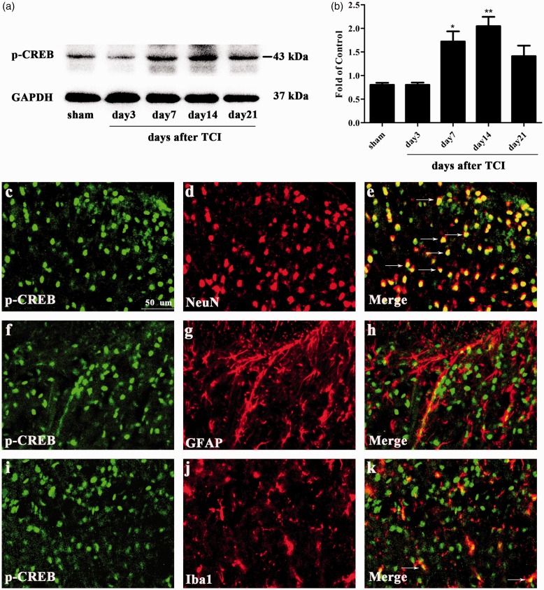
Figure 6.
Expression and cellular localization of phosphorylated-cyclic adenosine monophosphate response element-binding protein (p-CREB) in spinal cord dorsal horn of CIBP rats. (a) and (b) Western blot analysis showing the time course of p-CREB expression in sham and CIBP rats (*p < 0.05, **p < 0.01 compared with the sham group, n = 6 in each group). The fold change for the density of p-CREB was normalized to GAPDH for each sample, respectively. The fold change of p-CREB in the sham group was set at 1 for quantification. (c to k) Representative photomicrographs of p-CREB (green) double fluorescence labeling with NeuN (red) for neurons, GFAP (red) for astrocytes, and Iba1 (red) for microglia in the ipsilateral spinal cord at day 14 after TCI. Photomicrographs were taken from ipsilateral spinal cord dorsal horns of CIBP rats (n = 4 in each group). The results showed that p-CREB was predominantly co-expressed with neuron (yellow). TCI: tumor cell implantation; NeuN: neuronal nuclei; GAPDH: glyceraldehyde-3-phosphate dehydrogenase; GFAP: glial fibrillary acidic protein; Iba1: ionized calcium-binding adapter molecule 1; p-CREB:phosphorylated-cyclic adenosine monophosphate response element-binding protein.

