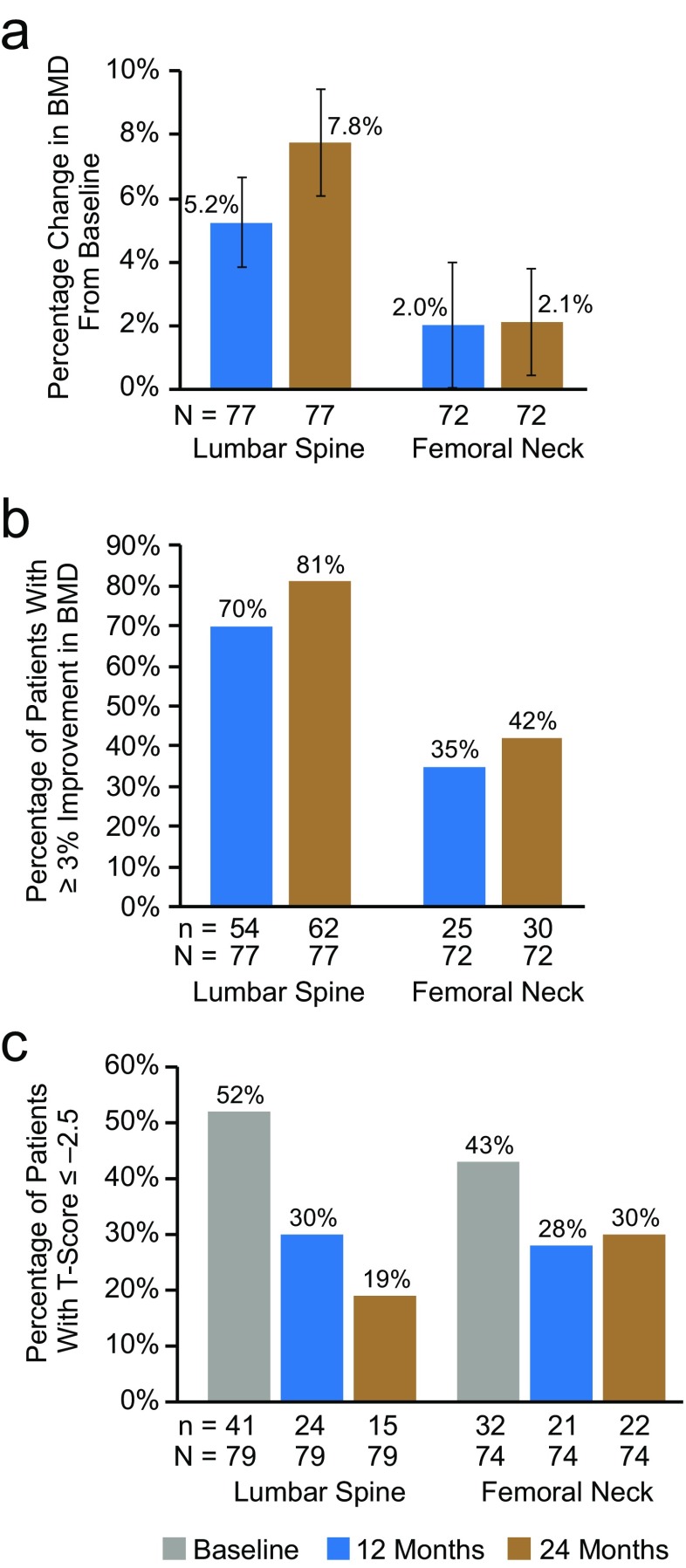
Fig. 3.
BMD at the lumbar spine and femoral neck in patients who were persistent at 24 months and who had a baseline, 12-month, and 24-month DXA scan. a Mean percentage change in BMD from baseline. Error bars represent 95% confidence intervals; 77 and 72 patients were evaluable at the lumbar spine and femoral neck, respectively. b Proportion of patients with ≥ 3% improvement in BMD at 12 and 24 months; 77 and 72 patients were evaluable at the lumbar spine and femoral neck, respectively. c Proportion of patients with T-scores ≤ − 2.5 at baseline, 12 months, and 24 months; 79 and 74 patients were evaluable at the lumbar spine and femoral neck, respectively. BMD bone mineral density; DXA dual-energy x-ray absorptiometry

