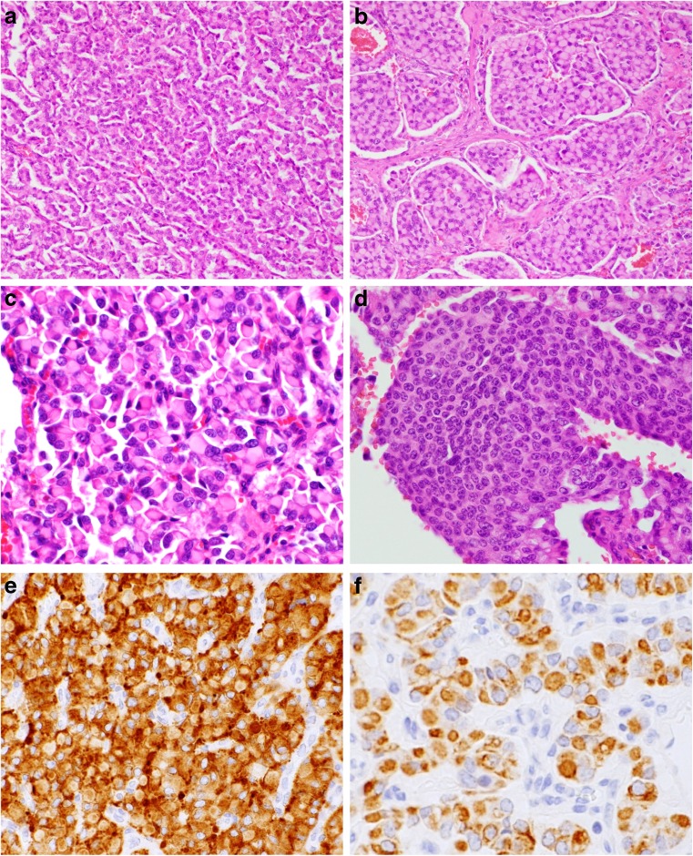
Fig. 2.
Histological findings for the tumor located in the pancreas head (a, c–f) and liver (b). a Tumor cells arranged in a cord fashion. b Tumor cells arranged in a nested fashion. c Tumor cells with abundant cytoplasm and densely eosinophilic globular inclusions that displaced the nuclei toward the periphery. d Uniform cells arranged in a nested fashion, demonstrating typical features of a pancreatic neuroendocrine tumor. e Tumor cells were positive for synaptophysin. f Immunohistochemically, intracytoplasmic globular inclusions were positive for AE1/AE3. Original magnification × 200 (a, b), × 400 (c–f)

