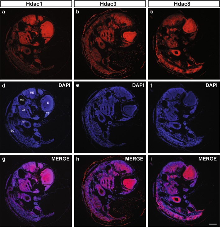
Fig. 2.
Ubiquitous expression of Class I Hdacs in N. furzeri embryos. Fluorescence immunohistochemistry (IHC) stainings of a Hdac1, b Hdac3, and c Hdac8 on sections from 15 day old embryos. Nuclei are counterstained with DAPI (d–f) and merged with signals obtained from class I Hdac-specific stainings (g–i). GI gills, GT gut tube, E eye, OV otic vesicle, RE rhombencephalon, SC spinal cord, TE telencephalon. Scale bar: 100 μm

