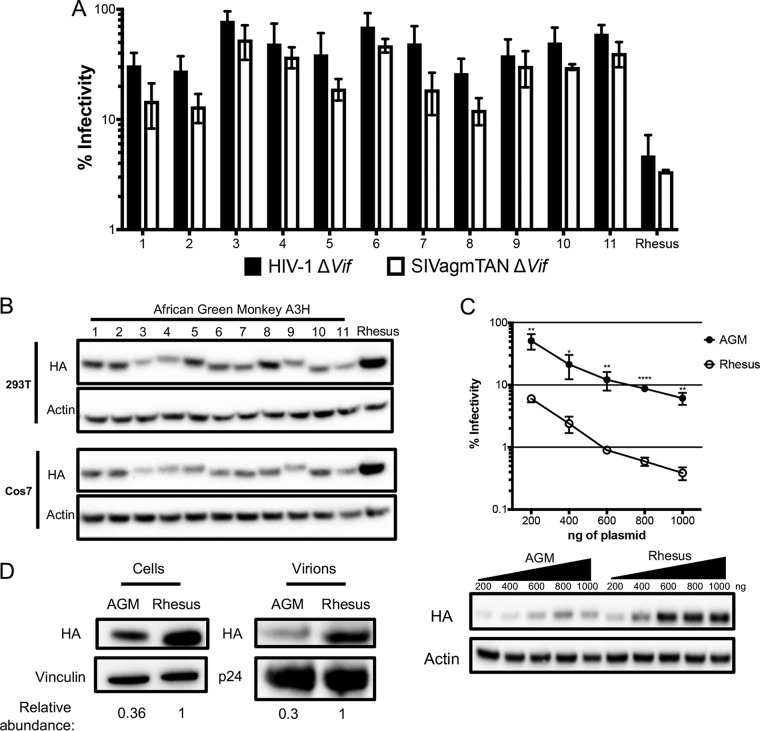
FIG 2.
Antiviral activity of A3H is lower in AGMs than in another Old World monkey. (A) Single-cycle infectivity assays were performed in the presence or absence of A3 proteins against HIVΔvif and SIVagmΔvif. Rhesus macaque was included as a positive control. Relative infection was normalized to viral infectivity in the absence of A3 proteins. Averages of three replicates, each with triplicate infections (±SEM), are shown. All the samples were statistically significantly different than rhesus macaque A3H, except AGM haplotype 7 against SIVagm.TAN (P = 0.0502), as measured by unpaired t tests. Furthermore, no significant differences in restriction between HIV and SIVagm.TAN within individual haplotypes were found. (B) Western blot analysis of HA-tagged AGM A3H protein expression in human (HEK293T) and AGM (Cos7) cell lines. The different-size bands for different AGM A3H haplotypes were reproducible. β-Actin is shown as a loading control. (C) (Top) Single-cycle infectivity assay of HIVΔvif in the presence of increasing amounts of A3H-expressing plasmids. AGM A3H haplotype 1 (black circles) and rhesus macaque A3H (open circles) were compared. Relative infection was normalized to viral infectivity in the absence of A3 proteins. Averages of three replicates, each with triplicate infections (±SEM), are shown. Statistical differences were determined by unpaired t tests: *, P ≤ 0.05; **, P ≤ 0.01; ****, P ≤ 0.0001. (Bottom) Western blot analysis of protein expression levels with the same amounts of plasmid as in the graph above. β-Actin is shown as a loading control. (D) Packaging of A3H proteins into virions analyzed by Western blotting. Relative abundances in cellular expression (left) and virion incorporation (right) were determined compared to rhesus macaque A3H.

