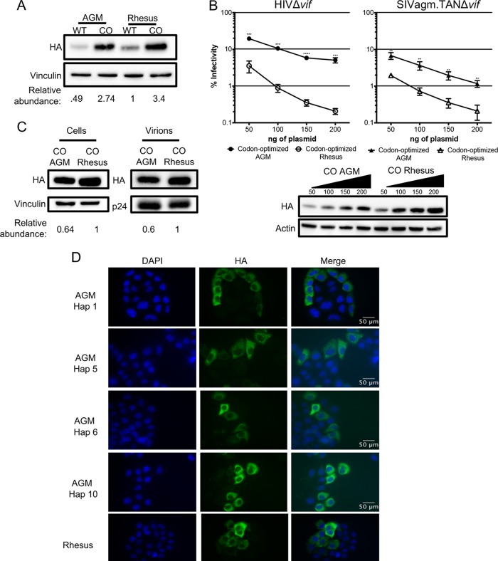
FIG 3.
Codon optimization increases protein expression and antiviral activity. (A) Western blot analysis for the expression of AGM A3H haplotype 1, codon-optimized haplotype 1 A3H, rhesus macaque A3H, and codon-optimized rhesus macaque A3H. Vinculin was used as a protein-loading control. Quantification was done relative to rhesus macaque A3H (normalized to 1). (B) (Top) Single-cycle infectivity assay of HIVΔvif and SIVagm.TANΔvif in the presence of increasing amounts of A3H plasmid comparing codon-optimized AGM haplotype 1 A3H (black) and codon-optimized rhesus macaque A3H (open). Relative infection was normalized to viral infectivity in the absence of A3 proteins. Averages of three replicates, each with triplicate infections (±SEM), are shown. Statistical differences were determined by unpaired t tests: **, P ≤ 0.01; ***, P ≤ 0.001; ****, P ≤ 0.0001. (Bottom) Western blot analysis of protein expression levels with amounts of plasmid added as for the graph above. β-Actin is shown as a loading control. (C) Packaging of A3H proteins into virions analyzed by Western blotting. Relative abundances in cellular expression (left) and virion incorporation (right) were determined compared to codon-optimized rhesus macaque A3H. (D) Subcellular localization of wild-type rhesus macaque and wild-type AGM A3H haplotypes (Hap) 1, 5, 6, and 10 in HeLa cells. A3H proteins were detected with an anti-HA antibody (green), and DAPI staining was used to detect the nucleus (blue). The images are representative of 135 total images over 3 replicates.

