FIG 3.

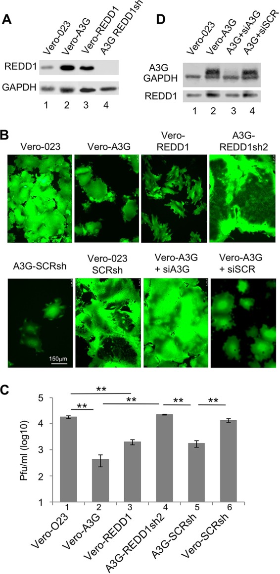
REDD1 expression and effects on MV replication. (A) The protein expression of REDD1 was assessed by Western blotting. Lysates of Vero-023 cells (lane 1), Vero-A3G cells (lane 2), REDD1 (F6gW-REDD1-Myc-DDK-tag)-transduced Vero (Vero-REDD1) cells (lane 3), and REDD1sh2-transduced Vero-A3G (REDD1sh2) cells (lane 4) were prepared, separated using 10% SDS-PAGE, and blotted on nitrocellulose, and the proteins were visualized with specific antibodies and the ECL system. (B) Vero-023 cells, Vero-A3G cells, Vero-REDD1 cells, Vero-A3G cells transduced with REDD1-specific shRNA-expressing vector (A3G-REDD1sh2), Vero-A3G cells transduced with nontargeted scrambled shRNA-expressing vector (A3G-SCRsh), Vero-023 cells transduced with nontargeted scrambled shRNA expressing vector (Vero-023-SCRsh), Vero-A3G cells transfected with A3G-specific siRNA, and Vero-A3G cells transfected with scrambled unspecific siRNA were infected with MV-eGFP at an MOI of 0.1 for 48 h. Representative photomicrographs of the eGFP fluorescence were taken to visualize the syncytium formation (magnification ×100; size bar, 150 μm). (C) Viral titers produced by these infected cells as indicated were determined 48 h after infection with MV. Mean viral titers from three independent experiments are shown (n = 3). Significances were calculated using Student's t test (**, P < 0.01). (D) Protein expression of A3G, GAPDH, and REDD1 in Vero-023 cells (lane 1), Vero-A3G cells (lane 2), Vero-A3G cells transfected with A3G-specific siRNA (lane 3), and Vero-A3G cells transfected with scrambled unspecific siRNA (lane 4) was determined by Western blotting.
