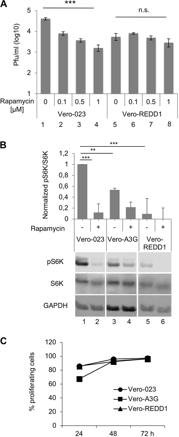
FIG 4.

Inhibition of MV replication by rapamycin and A3G and REDD1 effects on mTORC1 activity and proliferation of cells. (A) Vero-023 and REDD1-transduced Vero-023 (Vero-REDD1) cells were infected with MV-eGFP in the absence and presence of increasing concentrations of rapamycin as indicated. Viral titers were determined 48 h after infection of cells with MV at an MOI of 0.1. Because rapamycin was dissolved in dimethyl sulfoxide (DMSO), controls received the same concentration of DMSO (0.5%) as the rapamycin-treated cultures. The mean virus production from three independent experiments is shown (n = 3). Significances (comparison over four columns each) were calculated for each cell line using the one-way ANOVA test (***, P < 0.001; n.s., not significant). (B) Phosphorylation of S6K (Thr 389) and total S6K expression were determined by Western blotting using lysates of Vero-023 (bars 1 and 2), Vero-A3G (bars 3 and 4), and Vero-REDD1 (bars 5 and 6) cells in the absence and presence of 1 μM rapamycin. Controls received 0.5% DMSO. Western blots from three experiments were quantified and evaluated for statistical significance (Student's t test). (C) The proliferation of Vero-023, Vero-A3G, and Vero-REDD1 cells was determined by flow cytometry at 48 and 72 h. Cells were stained with the cell proliferation dye eFluor 670, and the percentage of cells with reduced signal intensity due to cell division was measured.
