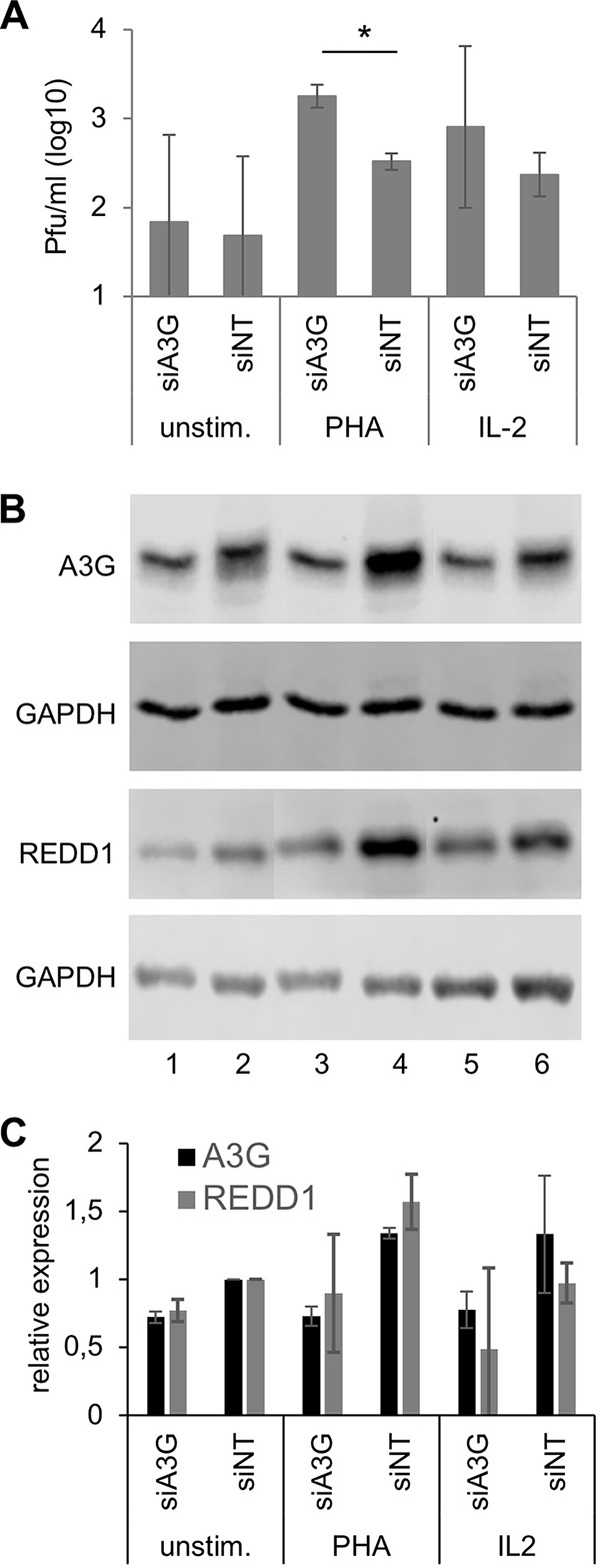
FIG 6.

Antiviral activity of A3G and effect on REDD1 expression in primary human PBL. Primary human PBL were cultivated in medium and transfected (nucleofected) with A3G-specific and nontargeted (NT) siRNAs. Cells were kept in medium (unstim.) or stimulated with 2.5 μg/ml PHA or 25 ng/ml IL-2. (A) At 24 h after stimulation, cells were infected with rMVIC323eGFP at an MOI of 0.1 for 48 h, and the newly synthesized virus was titrated using Vero-hSLAM cells. Mean values from three experiments with PBL from three independent donors are shown (significances were determined using Student's t test; *, P < 0.05). (B) Lysates were prepared from parallel uninfected cultures, and proteins were fractionated by SDS-PAGE and blotted on nitrocellulose membranes. A3G and REDD1 expression was visualized with specific antibodies and secondary antibodies on Western blots and the amount of protein on the blots controlled by staining with an antibody to GAPDH. A representative example is shown. (C) Quantification of A3G and REDD1 protein expression on Western blots from PBL from three independent blood donors. The relative protein levels of A3G and REDD1 were normalized and the expression in siNT-transfected PBL set to 1.
