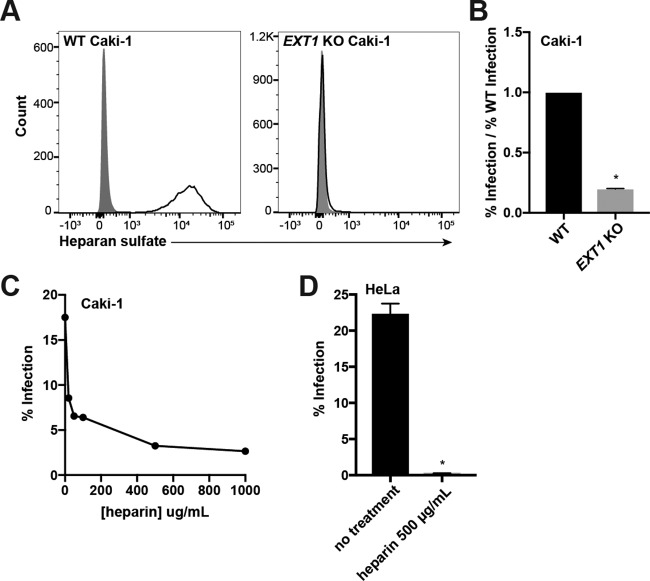
FIG 2.
Heparan sulfate interactions are required for infection of Caki-1 and HeLa cells. (A) WT and EXT1 KO Caki-1 cells were immunostained for surface heparan sulfate (HS) expression. Gray histograms represent isotype controls. (B) WT and EXT1 KO Caki-1 cells were infected with KSHV in duplicate, and infection rates were measured by flow cytometry. The infection rate of the KO was normalized to the average WT infection rate, and data were pooled from multiple experiments. (C) Filtered KSHV was preincubated with the indicated concentrations of soluble heparin at 37°C and then used to infect Caki-1 cells for 2 h at 37°C. Infection percentages were measured by flow cytometry at 2 days postinfection. (D) Filtered KSHV was preblocked with 500 μg/ml of heparin at 37°C and then used to infect WT HeLa cells in triplicate for 2 h at 37°C. The infection percentage was measured by flow cytometry at 2 days postinfection. *, P < 0.05.

