
FIG 5.
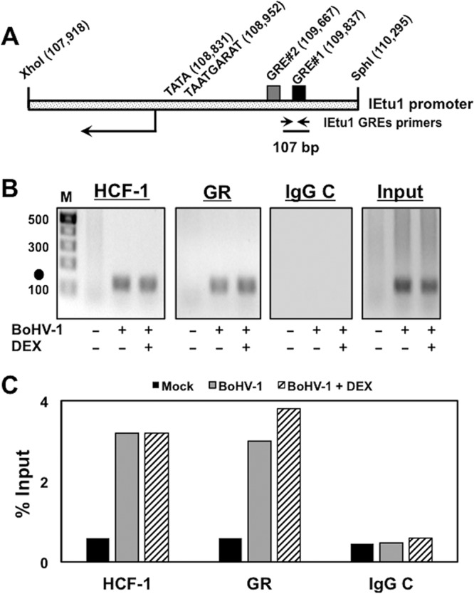
HCF-1 and GR occupy the BoHV-1 IEtu1 promoter in BoHV-1-infected cells. (A) Schematic of the IEtu1 promoter with the genomic locations of the TATA box, TAATGARAT core, GREs, and IEtu1 GRE ChIP primer pair. (B) CRIB cells were mock infected (−) or infected with BoHV-1 (+, MOI of 1) and treated with vehicle (−) or DEX (+) for 7 h. CRIB cells were transfected with the GR expression plasmid (1 μg plasmid) for 12 h prior to infection. HCF-1 and GR occupancy were assessed by ChIP assays using the DRR primer set. IgG, control; input, 10% of total DNA; M, DNA size markers (bp). The results are representative of three independent studies. Closed circles denote 107-bp PCR product. (C) The intensity of bands in panel B was quantified using a Bio-Rad ChemiDoc system.