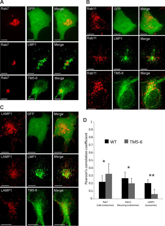
FIG 6.
TM5-6 exhibits altered colocalization with endolysosomal markers. HEK293 cells were cotransfected with GFP-tagged LMP1 constructs and RFP-tagged (A) Rab7, (B) Rab11, or (C) LAMP1. Live-cell confocal images were acquired 24 h posttransfection. The images were analyzed using Imaris software. (D) Colocalization was quantified using Pearson's correlation coefficient and graphed with standard errors of the mean (n ≥ 12 cells). Representative maximum-projection images are shown. *, P < 0.05; **, P < 0.001. Scale bars, 10 μm.

