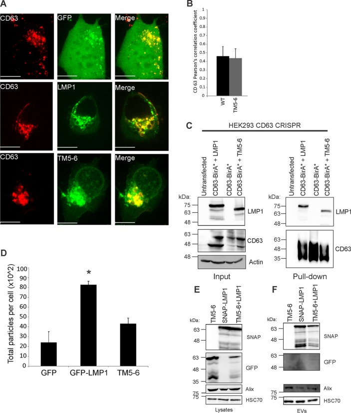
FIG 7.
TM5-6 colocalizes and associates with CD63. (A) HEK293 cells were cotransfected with GFP-tagged LMP1 constructs and CD63-RFP. Live-cell confocal images were acquired 24 h posttransfection. The images were analyzed using Imaris software. Representative maximum-projection images are shown. Scale bar, 10 μm. (B) Colocalization was quantified using Pearson's correlation coefficient and graphed with standard errors of the mean (n ≥ 12 cells). (C) CD63 knockout cells were cotransfected with CD63-BirA* plus WT LMP1 or CD63-BirA* plus TM5-6 or transfected with CD63-BirA* alone. Biotinylated protein complexes were isolated and separated by SDS-PAGE and analyzed by immunoblotting with LMP1 and CD63-specific antibodies. (D) EVs were harvested from HEK293 cells transiently transfected with GFP, GFP-LMP1, or TM5-6 and quantified by nanoparticle tracking. The data are represented as total numbers of particles harvested per cell. *, P < 0.05. (E and F) HEK293 cells were transfected with GFP-TM5-6, SNAP-LMP1 or cotransfected with GFP-TM5-6 and SNAP-LMP1. (E) Whole-cell and (F) EV lysates were separated by SDS-PAGE and analyzed by immunoblot analysis for GFP, SNAP, and common EV markers (Alix and HSC70).

