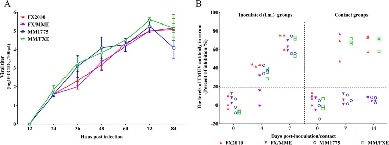
FIG 3.
Growth curves of parental and E gene substitution TMUVs on DF-1 cells and antibodies detected in contact and inoculated ducks with these viruses. (A) Replication of parental and E gene substitution TMUVs. DF-1 cells were infected by the parental and E gene substitution TMUVs at an MOI of 0.0001. Virus samples from the supernatant were collected at different time points and titrated on DF-1 cells. The data for virus titers indicate the means of the results of three repeats, and the error bars indicate standard errors of the means (*, P < 0.05; **, P < 0.01). (B) Relative levels of TMUV-specific antibodies detected in serum samples as a function of time after inoculation with the indicated virus. Serum was considered positive when the PI value was ≥18.4%.

