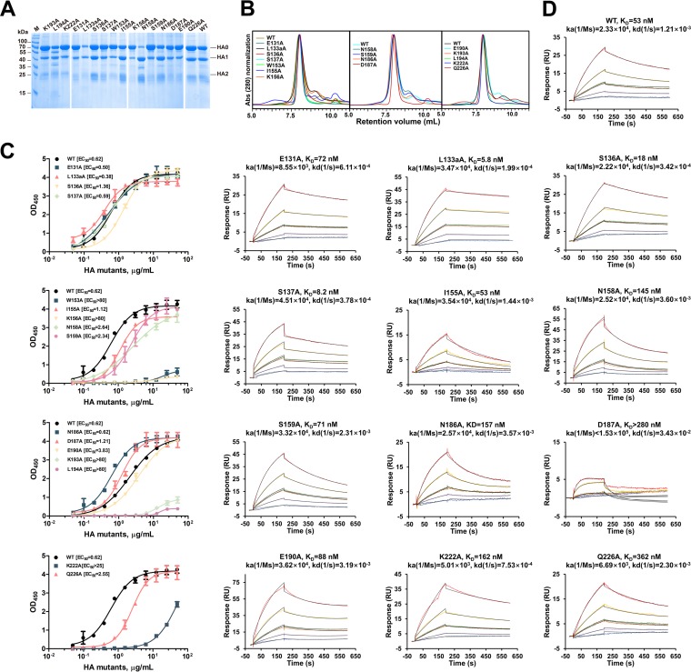
FIG 3.
Characterization of HA mutants with structure-based alanine substitutions in 13D4 binding sites. (A) SDS-PAGE analysis of the purified HA mutants. Notably, most of the N158A mutant proteins were cleaved into HA1 and HA2. (B) HPLC profiles of the purified HA mutants. The mutants were resolved as major components in HPLC curves with the same retention volume as WT HA, indicating the proteins exist mainly as trimers in solution. Abs, absorbance. (C) The interaction of HA and its mutants against MAb 13D4 was tested by double-antibody sandwich ELISA, and EC50s were calculated by sigmoid trend fitting and plotted as in Fig. 2F. OD450, OD at 450 nm. The error bars represent standard deviations of two repeats. (D) Binding curves of HA, HA mutants, and MAb 13D4 in SPR. A total of 5 concentrations (8.93, 17.9, 35.7, 71.4, and 143 nM) of HA and its mutants (serial dilutions) were injected onto a MAb 13D4-bound chip. The kinetic constants between MAb 13D4 and HA/HA mutants are reported.

