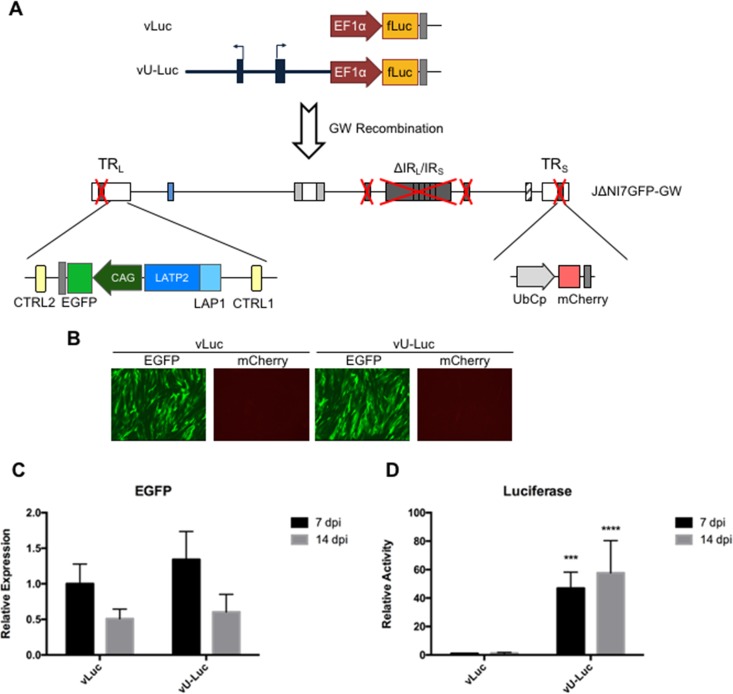
FIG 7.
vLuc and vU-Luc genome structures and transgene expression in HDFs. (A) Schematic drawing of the vLuc and vU-Luc genomes. A CAG-EGFP cassette was located in the LAT loci of both vectors. A GW cassette positioned in the intergenic region between UL50 and UL51 was used to introduce an EF1α promoter-luciferase (fLuc) expression cassette with or without upstream A2UCOE. (B) EGFP and mCherry fluorescence in vLuc- and vU-Luc-infected HDFs at 7 dpi. (C) EGFP mRNA levels of vLuc- or vU-Luc-infected HDFs at 7 and 14 dpi expressed relative to vLuc at 7 dpi (qRT-PCR data were normalized to viral gc in the same samples). (D) Luciferase activity in vLuc- or vU-Luc-infected HDFs at 7 and 14 dpi relative to vLuc at 7 dpi. The data are means and SD; ***, P ≤ 0.001, and ****, P ≤ 0.0001 compared to vLuc at the same time point (2-way ANOVA).

