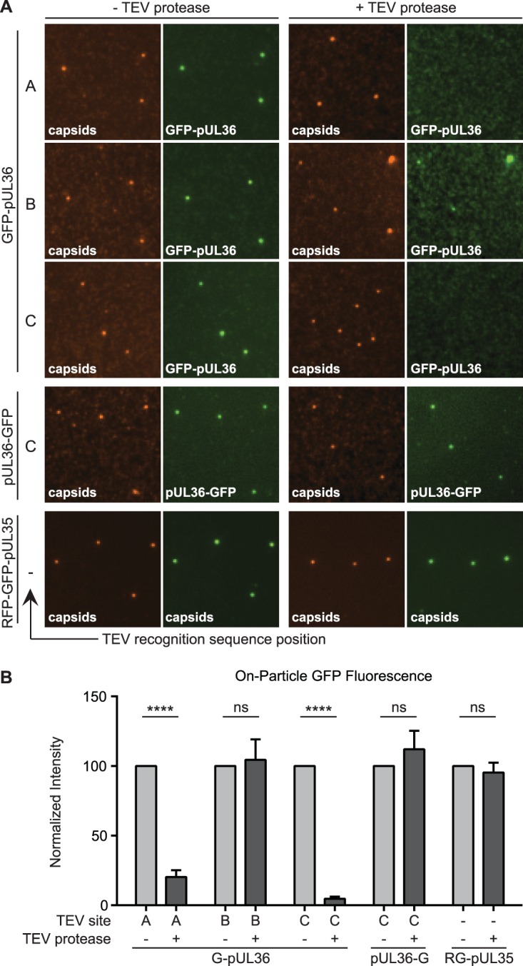
FIG 3.

Mapping regions of pUL36 that maintain stable attachment to capsids. (A) Representative images of dual-fluorescent extracellular virus particles that were detergent extracted and incubated in the absence (left two columns) or presence (right two columns) of TEV protease. The position of the TEV site within pUL36 (position A, B, or C) is denoted, with a dash indicating no TEV site. All viruses encode the pUL25/mCherry capsid tag, with the exception of the RFP-GFP-pUL35 dual-capsid-tagged virus. (B) Fluorescence intensities from single particles represented in panel A. Values are normalized to the cognate “− TEV protease” control and represent data from n ≥ 3 experiments. Error bars indicate standard errors of the means (****, P < 0.001).
