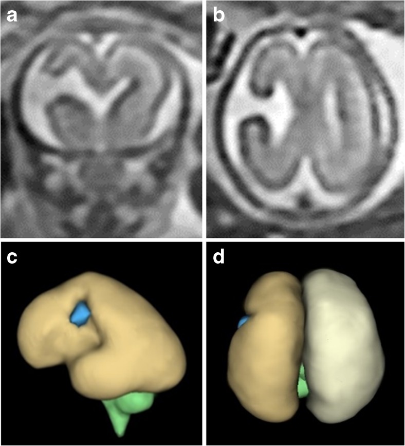
Fig. 11.
Unilateral schizencephaly (type 3) in a 21gw fetus. Coronal (a) and axial (b) ultrafast T2-weighted images show a widely spaced cleft in the right paracentral lobule. Those images are reversed to be consistent with the constructed models (c—left lateral and d—superior) of the brain constructed from a 3D steady-state acquisition. The septum pellucidum is absent but no other brain abnormality was shown

