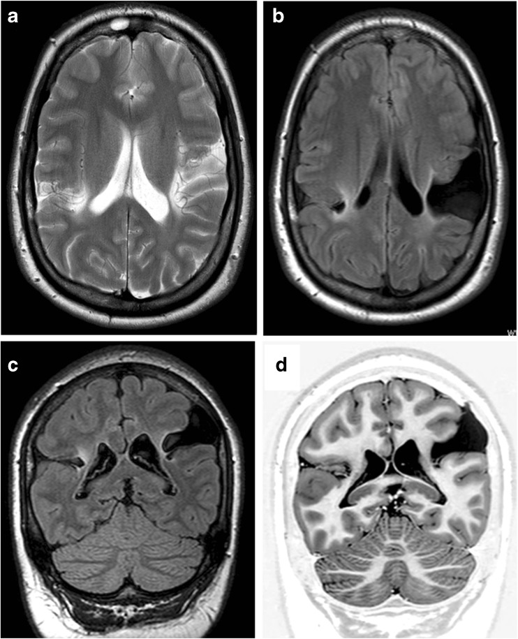
Fig. 2.
A child with porencephaly. MR imaging of an 8-year-old child with spastic quadriplegic cerebral palsy recognised in the first year of life. Axial T2-weighted (a), axial FLAIR (b), coronal FLAIR (c) and coronal inversion recovery (d) show bilateral clefts involving the paracentral lobules, which extend from the outer surface of the brain but do not quite reach the ventricular margin. Some of the white matter next to the clefts is gliotic and there is no evidence of normal, or abnormal, grey matter lining the clefts. These features indicate porencephaly rather than schizencephaly

