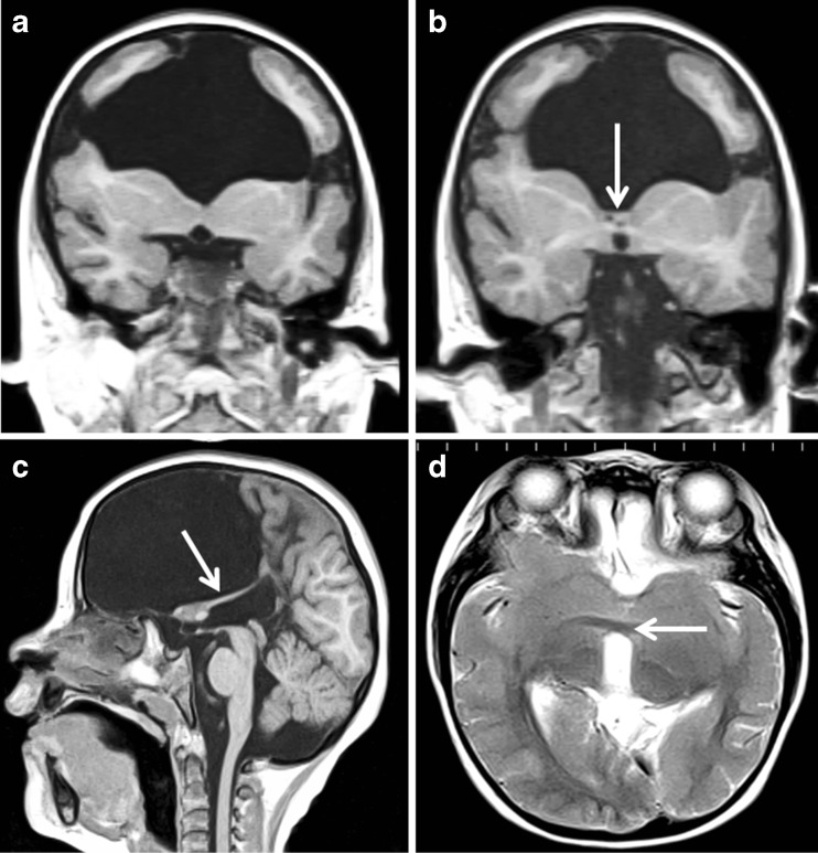
Fig. 8.
Bilateral schizencephaly (type 3) with multiple other brain abnormalities. MR imaging of a 5-year-old child with global developmental delay and epilepsy. Coronal reconstructions from T1 volume datasets (a, b) show bilateral schizencephaly (type 3) involving the inferior frontal gyri. Bilateral cortical formation abnormalities are present in the superior portions of the frontal lobes. The corpus callosum and septum pellucidum are absent and the fornices have an abnormal low position (arrowed on b and c). Axial T2-weighted image shows abnormal fusion of the nucleus accumbens septi and basal forebrain across the midline (arrowed on d) which possibly represents a form of septo-preoptic holoprosencephaly

