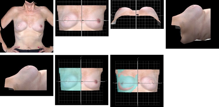
Fig. 3.
Protocol to measure breast symmetry, RMS Projection difference (RMS-PD). a AP view of torso, b gridlines placed onto image and cropped, c torso in craniocaudal view, d image rotated laterally, e image cropped at level of the anterior axillary line, f one-half of torso is selected, Image is copied and reflected in x = 0

