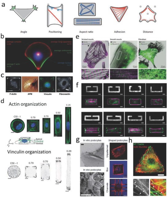
Figure 7.

The effect of geometrical cues on actin organization. a) Schematic image showing how geometry directs cytoskeleton organization. Reproduced with permission.71 Copyright 2012, Cell Press. b) Organization of polarity is governed by cell adhesive microenvironment. Reproduced with permission.72 Copyright 2006, National Academy of Sciences (United States). c) Organization of the stress fibers, FAs, and ECM within cells on a patterned square ECM island. Reproduced with permission.73 Copyright 2002, FASEB. d) Cell aspect ratio changes affect organization of actin stress fibers and FAs. Reproduced with permission.74 Copyright 2012, Nature Publishing Group. e) Actin, myosin II, and α‐actinin staining for different cell types. Reproduced with permission.75 Copyright 2015, Nature Publishing Group. f) Dissipation of elastic energy in severed stress fibers depends on fiber length. Reproduced with permission.76 Copyright 2017, National Academy of Sciences (United States). g) (Left) Scanning electron microscopy (SEM) images show in vivo podocytes with branched structure; (Right) F‐actin staining for cells cultured on glass, box, and microchannels. Reproduced with permission.77 Copyright 2017, Nature Publishing Group. h) Microtubule growth trajectories are correlated with F‐actin bundles controlled by cell geometry. Reproduced with permission.78 Copyright 2012, The Company of Biologists (United Kingdom).
