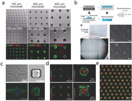
Figure 9.

Microwells in cell biology studies. a) ESCs cultured in PEG microwells with different diameters for 7 d. Reproduced with permission.[[qv: 50b]] Copyright 2009, National Academy of Sciences (United States). b) High‐throughput platform based PEG microwells for investigating single cell fate. Reproduced with permission.102 Copyright 2009, Royal Society of Chemistry (United Kingdom). c) Confocal images show cells cultured in PDMS microwells with different shapes. Reproduced with permission.103 Copyright 2007, Royal Society of Chemistry (United Kingdom). d) Controlling spatial organization of multiple cell types in microwells with certain 3D geometries. Reproduced with permission.104 Copyright 2012, Wiley. e) Microwells can be used for creating microparticle arrays with complex building blocks, green particles are assembled before red particles. Reproduced with permission.105 Copyright 2017, Nature Publishing Group.
