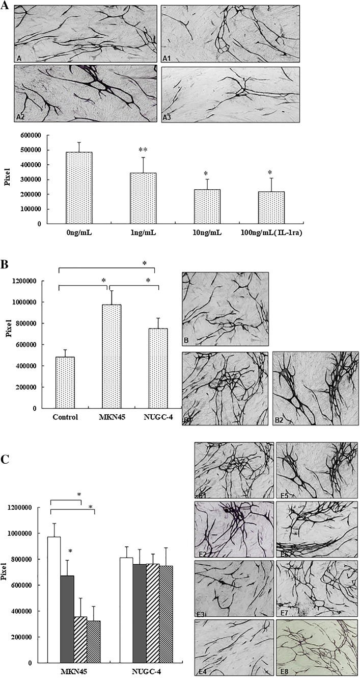
Fig. 5.
a Effect of IL-1RA on HUVEC tube formation. A, 0 ng/mL IL-1RA; A1, 1 ng/mL IL-1RA; A2, 10 ng/mL IL-1RA; A3, 100 ng/mL IL-1RA (×200). Columns Mean pixels of HUVEC tube formation area. Asterisks indicate significance at *P < 0.01 and **P < 0.05 vs. the control (0 ng/mL IL-1RA). b Effect of MKN45 and NUGC-4 on angiogenesis using the double chamber approach. Tube formation by HUVECs was significantly enhanced by co-culture with MKN45 cells compared with control or co-culture with NUGC-4. Asterisks indicate significant difference at *P < 0.01. B, Control; B1, co-culture with MKN45; B2, co-culture with NUGC-4. c Effect of IL-1RA on HUVEC tube formation in co-culture system. ×200 (0 ng/mL, white columns; 1 ng/mL, black columns; 10 ng/mL, left diagonal striped columns 10 ng/mL; 100 ng/mL, right diagonal striped columns). E1, Co-culture with MKN45; E2, with MKN45 + IL-1RA (1 ng/mL); E3, with MKN45 + rIL-1RA (10 ng/mL); E4, with MKN45 + rIL-1RA (100 ng/mL). E5, Co-culture with NUGC-4; E6, with NUGC-4 + IL-1RA (10 ng/mL); E7, with NUGC-4 + rIL-1RA (10 ng/mL); E8, with NUGC-4 + rIL-1RA (100 ng/mL). Asterisks indicate significant difference at *P < 0.01. Columns and whiskers Mean and SD

