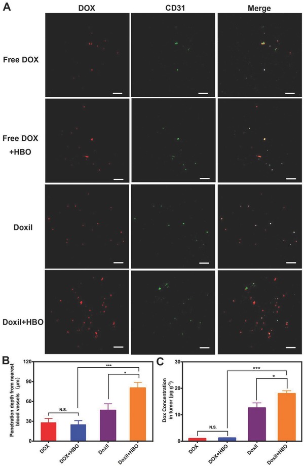
Figure 3.

In vivo drug penetration and tumor DOX accumulation. A) In vivo penetration of DOX into the tumors of H22‐bearing mice after intravenous injection of free DOX/Doxil at DOX dosage of 7 mg kg−1 with and without HBO therapy. The frozen tumor sections were observed at 24 h after injection using confocal microscopy. The blood vessels were stained by FITC‐CD31 antibody. The scale bar is 200 µm. B) The tumor penetration distance of DOX from the nearest blood vessel was determined with the simulated scatter diagrams method. C) The concentration of DOX in tumor tissue in H22‐bearing mice after intravenous injection of free DOX/Doxil at DOX dosage of 7 mg kg−1 with and without HBO therapy for 24 h. Data as mean ± S.E. (n = 5). *P < 0.05, ***P < 0.001. N.S. as not significant.
