Figure 2.
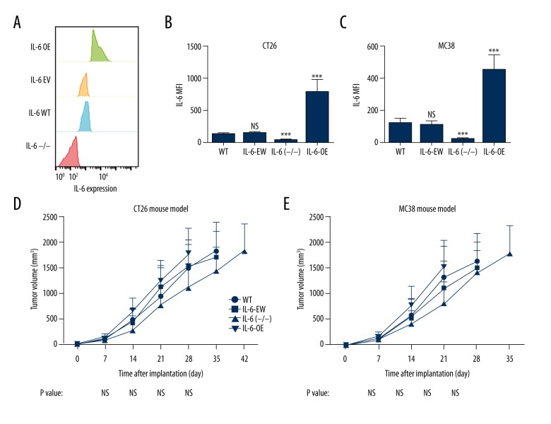
Role of IL-6 in tumor growth in CRC mouse models. (A) Expression of IL-6 was tested by flow cytometry in modified cells. (B, C) IL-6 expression values in modified cells. (D, E) Syngeneic mouse models were established using CT26 and MC38 cells with various levels of IL-6 expression. Tumors with IL-6 deficiency (IL-6 (−/−)) grew slightly slower than the tumors with IL-6 overexpression (IL-6-OE) and control groups, but no significant difference was observed. *** P value less than 0.001; WT – wild-type; EV – empty vector; OE – overexpression; MFI – mean fluorescence intensity; sample size of every experimental group was 10.

