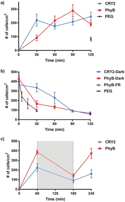
Figure 2.

a) Adhesion kinetics of CRY2‐MDA cells on CIBN‐functionalized substrates under blue light and PhyB‐MDA cells on PIF6‐functionalized substrates under red light. b) Reversion kinetics of light‐controlled cell–material interactions. First, CRY2‐MDA cells attached to CIBN‐functionalized substrates under blue light and PhyB‐MDA cells attached to PIF6‐functionalized substrates under red light for 1 h. Then, CRY2‐MDA cells were moved into the dark and PhyB‐MDA cells into the dark or under far‐red (FR) light. c) Switching of light‐controlled cell–material interactions. First, CRY2‐MDA and PhyB‐MDA cells attached to CIBN‐ or PIF6‐functionalized substrates, respectively, for 1 h under light, then were left in the dark (shaded in gray) for 2 h and placed again for 1 h under light. Blue and red light were used for CRY2‐MDA and PhyB‐MDA cells, respectively. The error bars are the standard error of three biological replicates each done in three technical replicates.
