Figure 1.
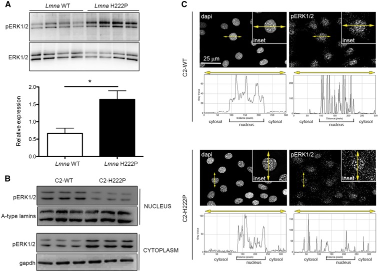
Increased ERK1/2 activation in hearts of LmnaH222P/H222P mice and in the cytoplasm of C2-H222P cells. (A) Immunoblots showing pERK1/2 and total ERK1/2 in hearts from LmnaH222P/H222P mice (H222P) and wild-type mice (WT). Data in bar graph below are represented as means±SEM (n=4; *P <0.05) from three independent repeats. (B) Immunoblots showing pERK1/2 and total ERK1/2 in extracts of nucleus and cytoplasm from C2-WT (n=3) and C2-H222P (n=3) cells. A-type lamins and gapdh are shown as loading controls. (C) Representative immuofluorescence micrographs of pERK1/2 staining of C2-WT and C2-H222P cells. Nuclei counter-stained with dapi are also shown. Insets show a higher magnification. Scan line graphs represent the intensity of pERK1/2 and dapi staining along the yellow arrow lines.

