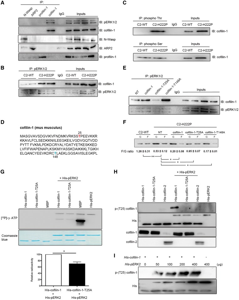
Figure 3.
Interaction between cofilin-1 and pERK1/2. (A) Immunoblot showing interaction of cofilin-1 and pERK12 in IP experiments. Proteins extracted from C2C12 cells were subjected to IP using antibodies against cofilin-1, N-Wasp, ARP2 or profilin-1. Proteins in immunoprecipitates were separated by SDS-PAGE and IB using antibodies against pERK1/2, cofilin-1, N-Wasp, ARP2 or profilin-1. Immunoglobulin G (IgG) was used as a negative control. Representative from three independent repeats. (B) Immunoblot showing interaction of cofilin-1 and pERK12 in IP experiments from C2-WT and C2-H222P cells. Proteins extracted from C2-WT and C2-H222P cells were subjected to IP using antibodies against pERK1/2. Proteins in immunoprecipitates were separated by SDS-PAGE and IB using antibodies against pERK1/2 and cofilin-1. IgG was used as a negative control. Representative from three independent repeats. (C) Proteins extracted from C2-WT and C2-H222P cells were subjected to IP using antibodies specific to phospho Thr or phospho Ser. The immunoprecipitates were separated by SDS-PAGE and IB using antibody against cofilin-1. IgG was used as a negative control. Representative from three independent repeats. (D) Amino acid sequence of murine cofilin-1 with highlighted threonine 25 (red) and threonine 148 (blue). (E) C2-H222P cells transfected or not transfected (NT) with plasmids encoding cofilin-1, cofilin-1-T25A or cofilin-1-T148A were subjected to IP using antibodies against pERK1/2. Proteins in immunoprecipitates were separated by SDS-PAGE and IB using antibodies against cofilin-1 or pERK1/2. IgG was used as a negative control. Representative from three independent repeats. (F) Immunoblot illustrating the effect of transfection with different cofilin-1 constructs on the amount of G-actin and F-actin and the calculated F/G actin ratio. Data are represented as means±SEM (n=3; *P <0.05, **P <0.005) from three independent repeats. (G) Representative immunoblot of in vitro kinase assay using recombinant histidine-tagged wild-type cofilin-1 (His-cofilin-1-WT) and cofilin-1-T25A (His-cofilin-T25A), without or with the addition of recombinant pERK2. MBP was used as a positive control. [32P]-γ-ATP indicates recombinant protein phosphorylation. Corresponding Coomassie blue-stained gel is shown below immunoblot. Data in bar graph below are means±SEM (n=3; *P<0.05) from three independent repeats. (H) Representative immunoblot of in vitro kinase assay using recombinant His-cofilin-1, His-cofilin-1-T25A and His-cofilin-2 without or with the addition of recombinant pERK2. Phosphorylated (Thr25) cofilin-1 was detected using a specific antibody. Representative from three independent repeats. (I) Representative immunoblot of in vitro kinase assay using recombinant cofilin-1 without or with the addition of recombinant pERK2 at increasing doses. Phosphorylated (Thr25) cofilin-1 was detected using the specific antibody. Representative from three independent repeats.

