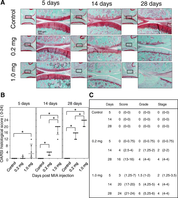
Fig. 5.
Articular cartilage changes after the MIA injection. a Representative images of Safranin-O staining of sagittal sections of the medial femoral and tibial condyles at each time point. b, c Osteoarthritis Research Society International histological scores were blindly evaluated by two independent researchers and data are presented in these panels. There were 6 samples at each time point. Four sections were randomly selected from each sample and the mean values were recorded. Median and quartile values in panel C are indicated. Asterisks indicate statistically significant differences. MIA, monoiodo-acetic acid

