Figure 1.

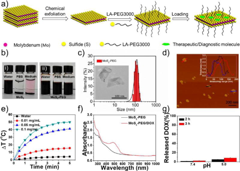
Synthesis and characterization of MoS2-based NSs. (a) Scheme of the exfoliation, PEGylation, and loading of MoS2 NSs with therapeutic/diagnostic molecules. (b) Colloidal stability of (i) MoS2 NSs and (ii) PEGylated MoS2 NSs in water, PBS, and cell culture medium after 48 h incubation. (c) DLS hydrodynamic size distribution of PEGylated MoS2 NSs. Inset: TEM image of PEGylated MoS2 NSs. (d) AFM image of PEGylated MoS2 NSs. The inset shows the thickness of PEGylated MoS2 NSs. (e) Photothermal heating curves of pure water and MoS2-PEG solutions with different concentrations (0.01, 0.05, and 0.1 mg/mL) under 808 nm laser irradiation at a power density of 1 W/cm2. (f) UV–vis-NIR absorbance spectra of PEGylated MoS2 NSs and PEGylated MoS2/DOX NSs. (g) The released DOX percentage from fluorescent NSs at pH 7.4 and 5.0 after 2 or 3 h incubation.
