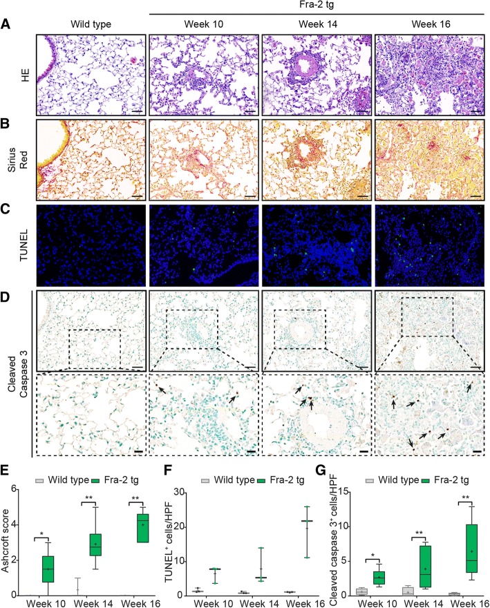
Fig. 3.
Time line of apoptosis in the fos-related antigen 2 (Fra-2) transgenic (tg) mouse model. Hematoxylin and eosin (HE) staining (× 20) (a); Sirius Red staining (collagen fibers identified by red staining; × 200) (b); TUNEL staining (× 200) (c); cleaved caspase 3 staining (× 200) (d), inlets show higher magnifications (×400), arrows highlight apoptotic cells; Ashcroft scores (e), semi-quantification of TUNEL+ cells (f) and semi-quantification of cleaved caspase 3+ cells (g): n = 3–4 (wild type) or n = 3–6 (Fra-2 tg). Data in box plots are median (line), mean (+) and minimum and maximum values: *p <0.05, **p < 0.01, Mann-Whitney U test. Scale bars 50 μm and 20 μm for inlets

