Fig. 6.
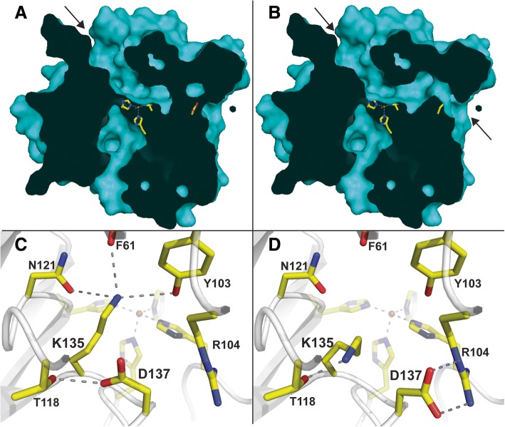
Active site cavities in PbLSD. a A surface slice of the derived crystal structure. The arrow is highlighting the classic stilbene substrate access cavity in the native PbLSD structure, from which product is expected to exit. b A surface slice of the derived crystal structure highlighting the tunnel cavity that can also exist within PbLSD by rotation of the side chains of amino acids Lys135 and Arg137, consistent with tunnel cavities in other carotenoid cleaving enzymes. Due to these images being produce by ‘slicing’, the side chain of Lys135 is not visible (its on the side of the slice that was cut off), but the side chain of Asp137 is visible blocking the channel in panel a and rotated away in panel b. Representations of the region surrounding Lys135 and Arg 137, highlighting the different molecular interactions observed are shown in (c) the native structure and (d) the native structure with Lys 135 and Arg 137 rotated to form a tunnel through the enzyme

