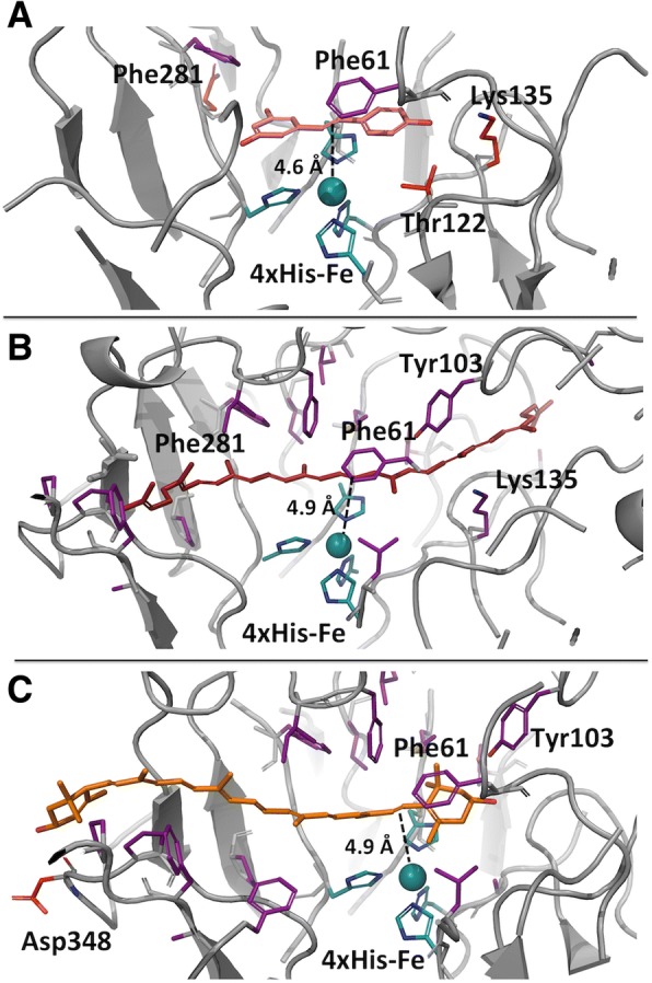
Fig. 7.

Virtual docking of molecules in the binding pocket of the crystal structure of enzyme PbLSD. A cartoon representation of the active site of PbLSD docked in proximity to the catalytic site 4-His-Fe subunit. a Resveratrol (b) lycopene and (c) lutein, all with the double bond most proximal to the catalytic center indicated by a dotted line (15–15′ and 7′-8′ respectively for lycopene and lutein). Potential pi-stacking and hydrogen bonding partners are labelled
