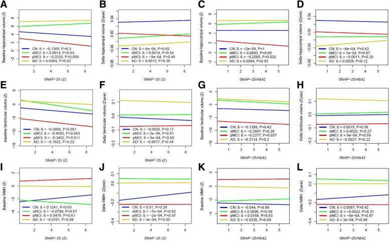
Fig. 8.
CSF SNAP-25 and SNAP-25/Aβ42 in relation to brain structure and WMH. Hippocampal volume, ventricular volume, and WMH at baseline (a, e, i) and over time (b, f, j) as a function of baseline CSF SNAP-25 in different diagnostic groups. Hippocampal volume, ventricular volume, and WMH at baseline (c, g, k) and over time (d, h, l) as a function of baseline SNAP-25/Aβ42 in different diagnostic groups. Biomarker levels and ratios are standardized to z scores. Aβ amyloid-β, AD Alzheimer’s disease, CN cognitively normal, pMCI progressive mild cognitive impairment, sMCI stable mild cognitive impairment, SNAP-25 synaptosomal-associated protein 25, WMH white matter hyperintensity

