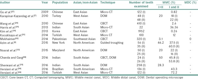
Table 2.
Prevalence of middle mesial and middle distal canals in mandibular first and second molars after introduction of three-dimensional imaging

Prevalence of middle mesial and middle distal canals in mandibular first and second molars after introduction of three-dimensional imaging
