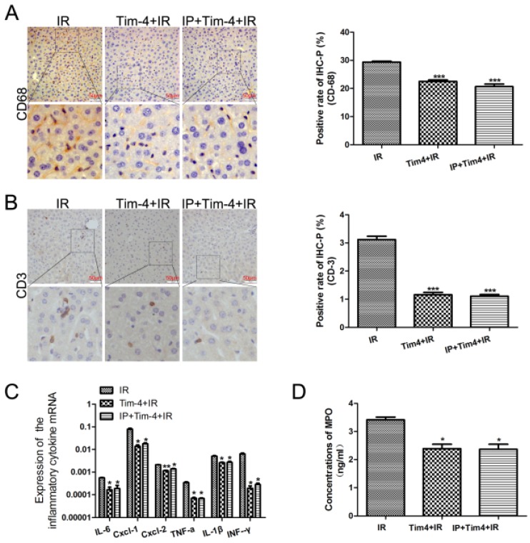
Figure 3.
Changes in inflammatory cells and cytokines in in mice subjected to IRI with or without IPC and TIM4 blocking. (A-B) CD68- and CD3-positive cells on immunohistochemical analysis (***P < 0.001 vs. IRI; n = 3 for each experiment). (C) Detection of IL-6, CXCL-1, CXCL-2, TNF-a, IL-1β, and IFN-γ using quantitative polymerase chain reaction assays. (*P < 0.05, **P < 0.01 vs IR). (D) MPO concentration determined using enzyme-linked immunosorbent assay (*P < 0.05 vs IR).

