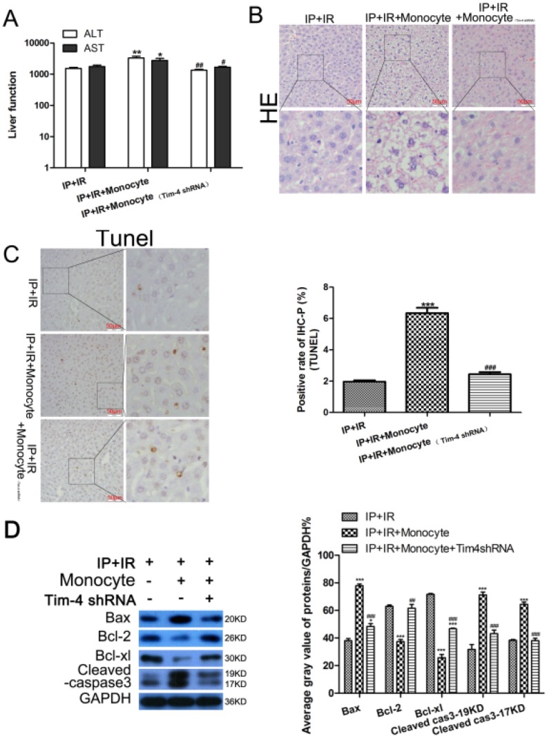
Figure 4.
Changes in serum transaminase levels, liver histology, and apoptosis-related proteins in mice subjected to IRI and ischemic preconditioning (IPC) with or without transfusion of activated monocytes and TIM4 blocking. (A) ALT and AST levels determined using enzyme-linked immunosorbent assay (*P < 0.05, **P < 0.01 vs. IPC+IRI; #P < 0.05, ##P < 0.01 vs. IPC+IRI+monocyte). (B and C) Liver histology examined using hematoxylin and eosin (HE) staining, and cell apoptosis examined using TUNEL assay (magnification, ×400; ***P < 0.001 vs. IPC+IRI; ###P < 0.001 vs. IPC+IRI+monocyte; n = 3 for each experiment). (D) Western blot analysis of apoptosis proteins expression (***P < 0.001 vs. IPC+IRI; ##P < 0.01 vs. IPC+IRI+monocyte; n = 3 for each experiment).

