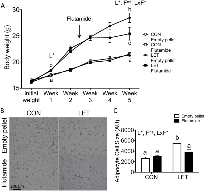
Figure 6.
Flutamide treatment reduced body weight and adipocyte cell size in LET females. (A) Body weights were measured weekly throughout the study; n = 9 to 10 per group. LET females weighed significantly more than CON after 1 week of LET, and flutamide-treated LET females weighed significantly less compared with LET mice that received empty pellets after 2.5 weeks of flutamide treatment. (B) Representative images of hematoxylin and eosin (H&E)–stained parametrial adipose tissue from CON and LET female mice that received empty pellet or flutamide treatment; n = 5 to 7 per group. (C) Quantification of mean adipocyte cell size from H&E-stained sections of parametrial adipose tissue; n = 5 to 7 per group. Images were quantified using ImageJ Fiji software with the Adiposoft plugin. Data were analyzed by two-way ANOVA with results indicated as follows: L, LET; F, flutamide; LxF, LET × flutamide interaction; *, significant effect; ns, no significant effect. Different letters indicate statistically significant differences between groups by Tukey post hoc analysis.

