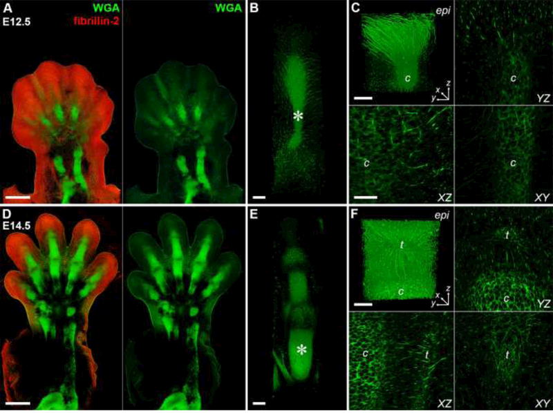
Fig. 4. Multiscale imaging of the musculoskeletal system in the developing forelimb.

E12.5 (A – C) and E14.5 (D – F) forelimbs were decellularized and stained with WGA (green) and an antibody against fibrillin-2 (FBN2, red). Dorsal view. (A, D) Cartilage elements of the autopod and zeugopod were easily resolved with WGA. FBN2 became more restricted with time. 10×, bar = 500 μm. (A: 2471 × 3599 × 600 μm; D: 2799 × 4733 × 450 μm; x × y × z) (B, E) 3D rendering of digits showed a network of WGA+ fibrils radiating from the cartilage as well as staining of joint tissues and the extensor tendons. 25×, bar = 100 μm. (381 × 1364 × 289 μm). (C, F) Higher magnification of digits in B, E (*) revealed WGA+ fibrils extended from dorsal epidermis (epi) to cartilage (c) at E12.5 and at E14.5 fibrils appeared to connect the extensor tendons (t) to the epidermis and cartilage. 40×, bars = 50 μm, (213 × 213 × 227 μm). See Movies 1 – 4.
