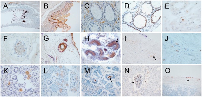
FIGURE 1.
Photomicrographs of autopsy sections immunohistochemically stained for pathological Asyn by the 2 compared methods illustrating common types of non-specific staining. Specific versus non-specific staining morphologies have been defined by their neuronal or non-neuronal appearance and empirically by their differential presence in normal control subjects versus subjects with autopsy-confirmed PD or other Lewy body diseases. Immunoperoxidase reaction product is brown, the counterstain is blue. Panels (A–E) are of sigmoid colon, (F–J) of skin from scalp, and (K–O) of submandibular gland. Most staining features were seen with both staining methods. (A, B) Sigmoid colon stained with the 5C12 method (A) and the Nantes method (B), showing non-specific DAB deposition on lumenal contents and within surface epithelial cells. (C) Mucosa of sigmoid colon, depicting frequent melanin-containing macrophages (melanosis coli) within the lamina propria, a relatively common non-specific finding (5C12 method). (D) Mucosa showing non-specific staining of epithelial cell nuclei; melanin-containing macrophages are also present in the lamina propria (5C12 method). (E) Macrophages non-specifically stained within the submucosa (5C12 method). (F, G) Non-specific staining of hair follicles ([F] with the 5C12 method, [G] with the Nantes method). Also seen in (G) are non-specifically stained melanocytes in the epidermis. (H) Sweat glands with non-specific staining of lumenal surfaces (arrow points to example; 5C12 method). (I) Collagen fibers (arrow points to an example) in the dermis, non-specifically taking up DAB (Nantes method). (J) Macrophages in the dermis with non-specific staining of cytoplasmic contents (5C12 method). (K, L) Diffuse, non-specific staining of cytoplasm of serous epithelial cells of submandibular gland ([K] with Nantes method, [L] with 5C12 method). (M) Non-specific staining of lumenal contents (arrow) of a duct within submandibular gland (5C12 method). (N) Non-specific DAB deposition on secretion (arrow) within a submandibular gland duct (5C12 method). (O) Non-specific DAB deposition along the edge of a submandibular gland biopsy (5C12 method).

