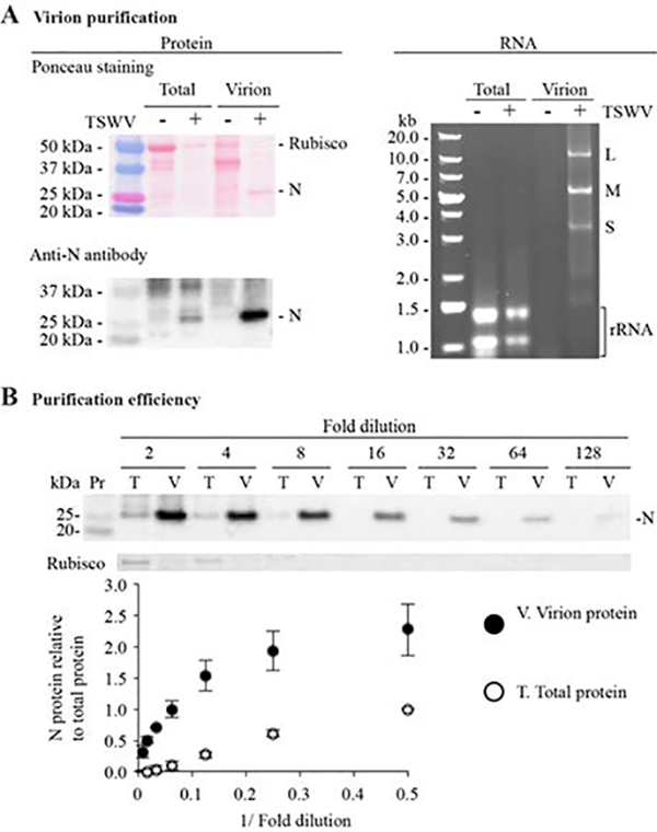
Figure 2.

Tomato spotted wilt virus (TSWV) virion purification and genomic RNA extraction. Total and virion protein and RNA extracted from 20 g mock-inoculated (−) or TSWV-infected (+) Nicotiana benthamiana plants at 21 days post inoculation. A. Virion purification. Left panel: representative blot stained with Ponceau S solution and an immunoblot showing nucleocapsid protein (N). Precision Plus Dual Color (161–0374; Bio-Rad) was used as a size marker. Right panel: 1% agarose gel electrophoresis analysis of total and virion RNA. GeneRuler 1-kb Plus DNA ladder (SM1333; Thermo Fisher Scientific) was used as a marker. Positions of the large (L), medium (M), and small (S) TSWV genomic RNAs and ribosomal RNA (rRNA) are indicated. B. Virion purification efficiency evaluated by a dilution curve. The chart shows signal intensities of the N protein bands measured with ImageJ. Values are reported as averages ± standard errors of four biological replicates expressed relative to the two-fold dilution of the total protein sample.
