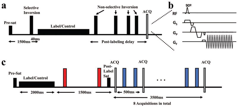
Figure 2.
Schematics of pulse sequences used in this study. (a) A conventional ASL sequence with a five-inversion background-suppression. (b) WEPCAST MRI sequence with flow-sensitive bipolar gradient added in the acquisition module. (c) Look-Locker WEPCAST MRI with background-suppression at all PLDs. The red pulses are vessel suppression pulses while the blue ones are tissue suppression pulses.

